Expertise in Aortic Aneurysms
-
What are the symptoms of an aortic aneurysm?
-
Are you at risk for an aortic aneurysm?
-
How do you treat an aortic aneurysm?
An internationally-recognized expert and leader in aortic aneurysm disease is here to help you!
Dr. Grayson Wheatley
Director of Aortic and Endovascular Surgery
Temple University School of Medicine
expertise. quality care. innovative therapies.
Case Study #1
Endovascular Repair of a Thoracic Aortic Aneurysm
Grayson H. Wheatley, MD, FACS
Patient History
An 84-year-old female presented to her primary care physician with complaints of a cold for a week. She was already being treated for the following medical conditions:
-
High blood pressure (hypertension)
-
Congestive heart failure
-
Coronary artery disease
-
Abnormal heart beat (atrial fibrillation)
-
Abnormal heart valve (mitral valve regurgitation)
-
Pulmonary hypertension
Diagnosis
A chest X-ray demonstrated a widening of the aorta in the chest. [image #1] To further clarify the diagnosis, a CT or CAT scan showed a 5.9cm thoracic aortic aneurysm (TAA). [image #2]
3D reconstructions of the CT scan images were used to evaluate the aortic aneurysm. [images #3 and #4]. This type of reconstruction is valuable in planning for treatment. Also, the abdominal aorta is evaluated to eliminate the possibility of another aortic aneurysm and to determine if the blood vessels in the thigh region (femoral arteries) are large enough to allow introduction of the thoracic aortic stent-graft. [image #5]
Treatment
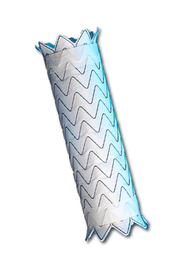
The patient underwent successful treatment of the aortic aneurysm using a thoracic aortic stent-graft, or endoluminal graft. [image #6]
The TEVAR procedure (Thoracic Endovascular Aneurysm Repair) is a minimally-invasive procedure performed in a special operating room, called a Hybrid OR. This room contains special X-ray equipment which allows the surgeon and team to carefully insert the medical device into an artery in the thigh-region (femoral artery) and guide the aortic-stent inside the aorta and deploy within the aneurysm sac.
Follow-up
After the TEVAR procedure, a follow-up CT scan was performed to ensure that the aneurysm was completely treated. [image #7] Following the successful procedure, she returned home after 3 days in the hospital and resumed all of her normal activities after 3 weeks of recovery at home.
Key Points
-
Aortic aneurysms often have no associated symptoms.
-
A CT scan is needed for diagnosis.
-
Special reconstructions of the CT scan are essential for planning treatment.
-
The TEVAR procedure is an endovascular procedure with an aortic stent-graft which allows for a more rapid recovery than traditional open surgery.
 1. Aortic aneurysm on chest X-rayThis chest X-ray is abnormal and demonstrates a widening of the heart shadow, called the mediastinal silhouette. This signifies that there is an enlargement of the aorta in the chest, called a thoracic aortic aneurysm. |  2. CT scan of the chestThis CT or CAT scan image demonstrates an aortic aneurysm in the chest which is behind the heart and next to the spine. The aortic aneurysm measures 5.9cm in maximal diameter. |  3. Chest CT scan 3D reconstructionThis 3D reconstruction image of the CT scan shows the aortic aneurysm as a localized widening of the aorta in the chest. The red color represents the normal aorta and the yellow color represents cholesterol which has built-up in the aortic wall contributing the aortic aneurysm (atherosclerosis). |
|---|---|---|
 4. Chest CT scan 3D reconstructionAnother 3D reconstruction image of the CT scan which shows the aortic aneurysm as a localized bulge in the chest. The red color represents the normal aorta and the yellow color represents cholesterol which has built-up in the aortic wall contributing the aortic aneurysm (atherosclerosis). |  5. Abdominal CT scan reconstructionIn order to evaluate a patient for an endovascular procedure, called TEVAR (Thoracic Endovascular Aneurysm Repair), a CT scan of the abdomen is needed to assess if the entry vessels in the thigh region are large enough to allow introduction of the aortic stent-graft. The white color represents calcium which has built-up in the wall of the aorta. |  6. Thoracic Aortic Stent-GraftThe TEVAR procedure is an endovascular procedure which avoids opening the chest and uses an aortic stent-graft, or endoluminal graft, and X-ray guidance to treat the aortic aneurysm in the chest. |
 7. Chest CT scan after aortic stentFollowing the TEVAR procedure, a CT scan of the chest shows the aortic stent-graft functioning properly and the aortic aneurysm is successful treated. |

